The LIMA global hyperspectral microscope uses excitation based on a widely tunable excitation source spanning 400 nm to 1620 nm, allowing for unique darkfield and brightfield imaging.
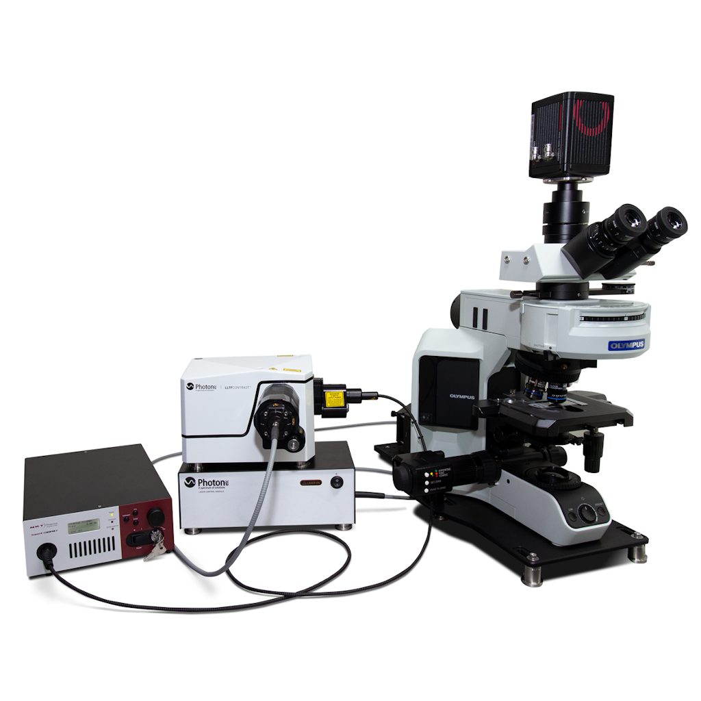
Photon etc.’s filtering technology illuminates the full field of view of a research-grade microscope with a continuously tunable monochromatic, high power density laser light. The system provides a high spectral resolution in the VIS, NIR, and SWIR combined to a near-diffraction-limited spatial resolution. Ideal for darkfield, PLE, or standard brightfield reflectance and transmittance imaging, it can map the full spectral response of a sample much faster than point-by-point or line-scan-based systems.
Three models of the LIMA are available all offer sub micron spatial resolution over the spectral range of operation.
The available models are:
LIMA VIS 400-1000nm, 1.5 – 2.5 nm spectral resolution. Detection via sCMOS (optionally EMCCD or CCD)
LIMA SWIR 900-1620 nm, 3.0 – 5.0 nm spectral resolution. Detection is via Photon etc. InGaAs camera (ZephIR 1.7 or Alizé 1.7)
LIMA eSWIR 1000-2500 nm, <5.0 nm spectral resolution. Detection is via Photon etc. HgCdTe camera ZephIR 2.7
VIS-SWIR Model | eSWIR | ||
---|---|---|---|
VIS | SWIR | ||
Excitation Spectral Range | 400-1000 nm | 900-1620nm | 1000-2500nm |
Excitation Spectral Resolution (FWHM) | 1.5-2.5nm | 3.0-5.0nm | <5.0nm |
Excitation Wavelength | Continuously Tunable | ||
Out-of-band Rejection | <-60dB @ ±40nm | < -60dB @ ±80nm | < -60dB @ ±80nm |
TLS Output Power | 2 to 4 mW | 3 to 9 mW | 0.5 to 8 mW |
Illumination | High efficiency homogenous illumination | ||
Dia or Epi Illumination; Brightfield; Darkfield (Oil and Dry) | |||
Spatial Resolution | Sub-micron – limited by the microscope objective NA | ||
Camera | sCMOS (Optionally CCD, EMCCD) |
Photon etc. InGaAs camera (ZephIR™ 1.7 or Alizé™ 1.7) | Photon etc. HgCdTe camera (ZephIR™ 2.5)M |
Microscope | Upright or inverted, scientific grade | ||
Wavelength Absolute Accuracy | FWHM/8 | ||
Scanning Speed | 20 to 50ms stabilization time (for step sizes 0.01 to 10 nm) | ||
X, Y Travel Range | 76 mm x 52 mm (with a manual stage) | ||
Z stage resolution | 100 nm | ||
White Light Illumination | Diascopic, episcopic, Hg, halogen | ||
Prepocessing | Spatial filtering, statistical tools, spectrum extraction, data normalization, spectral calibration, overlay, central position map, etc. | ||
Hyperspectral Data Format | HDF5, FITS | ||
Software | PC (Windows10 – 64-bits) with PHySpec™ control and analysis software (computer included) | ||
Dimensions | Microscope: 31 cm x 85 cm x 82 cm Supercontinuum laser: Up to 44 cm x 25.1 cm x 40 cm Tunable filter: Up to 30 cm x 23 cm x17.5 cm | ||
Combined: Less than 105 cm x 85 cm x 82 cm | |||
Weight | ≈ 80 Kg (Gross 220 lbs ; Net 180 lbs) | ||
Power Requirement | 120 VAC / 12 A / 60 Hz 230 VAC / 12 A / 50 Hz | ||
OPTIONS & ACCESSORIES | |||
Objectives magnifcation: 20X, 40X, 50X, 60X, 100X (5X and 10X available on demand) | |||
Motorized stage: 100 mm x 100 mm travel, 22 nm resolution | |||
Filter wheel: up to 6 band-pass filters | |||
Second camera port | |||
High resolution module: VIS < 0.5nm ; SWIR < 1.0nm | |||
Hyperspectral filter in detection (2D spectroscopic micro-imaging) | |||
Polarization control (polarizer & analyser) | |||
Optical table with passive anti-vibration isolation recommended: 900 x 1800 x 60 mm (36 x 72 x 2.4 inches) or 900 x 900 x 60 mm (36 x 36 x 2.4 inches) next to 900 x 900 mm (36 x 36 inches) standard table |
Dark Field Hyperspectral Imaging
Solar cell characterisation
Nanomaterial characterisation
Semiconductor quality control