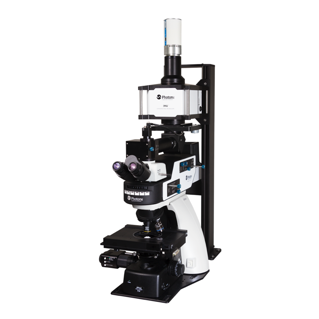
Photon etc.‘s IMA Global hyperspectral microscope delivers spectral and spatial information in the VIS, NIR and/or SWIR spectral ranges (400 nm – 1620 nm).
Photon etc.‘s IMA Global hyperspectral microscope delivers spectral and spatial information in the VIS, NIR and/or SWIR spectral ranges (400 nm – 1620 nm).
The IMA Global Hyperspectral Microscope system produces large maps (up to 1 mm x 1 mm and even more) of photoluminescence, fluorescence and electroluminescence, reflectance, and transmittance. Based on high throughput global-imaging filters, IMA is faster and more efficient than standard point-by-point or line-scan based systems. IMA is designed for both life sciences and materials science
Two models of the IMA can be offered:
IMA VIS that operates over 400-1000nm with <2nm spectral resolution (FWHM) and with sub nm resolution. Detection is via CCD, EMCCD or sCMOS camera
IMA SWIR that operates over 900-1620 nm with <4nm spectral resolution and with sub nm resolution. Detection is via Photon etc. InGaAs camera (ZephIR 1.7 or Alizé 1.7)
Quick mapping
High spatial and spectral resolution
Complete system (source, microscope, camera, filter, software)
Straight or inverted microscope
Non-destructive analysis
Custom specifications available
Darkfield and electroluminescence imaging option
| Vis-SWIR Model 400 – 1620 nm |
|||
|---|---|---|---|
| Spectral Range | VIS 400-1000nm |
SWIR 900-1620nm |
|
| Spectral Resolutions (FWHM) | <2 nm | <4 nm | |
| Spectral Channels | Continuously Tunable | ||
| Spatial Resolution | Sub-micron- limited by the microscope objective NA | ||
| Camera | CCD, EMCCD, sCMOS | Photon etc. InGaAs camera (ZephIR™ 1.7 or Alizé™ 1.7) | |
| Excitation Wavelengths (Up to 3 lasers) |
405, 447, 532, 561, 660, 730, 785, 808 nm (other wavelengths available upon request) |
||
| Microscope | Upright or inverted, scientific grade | ||
| Wavelength absolute accuracy | FWHM/8</td | ||
| Maximum Scanning Speed | 150 ms per Wavelength | ||
| X, Y Travel Range | 76mm x 52mm (with a manual stage) | ||
| Z Stage Resolution | 100nm | ||
| White Light Illumination | Diascopic, episcopic, Hg, halogen | ||
| Illumination Options | Epifluorescence module, darkfield module (oil or dry) | ||
| Video Mode | Megapixel camera for sample visualization | ||
| Preprocessing | Spatial filtering, statistical tools, spectrum extraction, data normalization, spectral calibration, overlay, central position map, etc. | ||
| Hyperspectral Data Format | HDF5, FITS | ||
| Software | PC (Windows10 – 64-bits) with PHySpec™ control and analysis software (computer included) | ||
| Dimensions | ≈ 150 cm x 85 cm x 82 cm | ||
| Weight | ≈ 80 kg | ||
| Power Requirement | 120 VAC / 12A / 60Hz 230 VAC / 12A / 50Hz |
||
| OPTIONS AND ACCESSORIES | |||
| Objectives magnification: 10X, 20X, 40X, 50X, 60X, 100X | |||
| Spectral range extension (e.g. UV Option with FWHM=10 nm) | |||
| Motorized stage: 100 mm x 100 mm travel, 22 nm resolution | |||
| Filter wheel: up to 6 band-pass filters | |||
| Electroluminescence module | |||
| Second camera port | |||
| Absolute photometric calibration | |||
| High resolution module: 900 – 1620 nm FWHM < 1 nm | |||
| *Optical table with passive anti-vibration isolation recommended: 900 x 1800 x 60 mm (36 x 72 x 2.4 inches) or 900 x 900 x 60 mm (36 x 36 x 2.4 inches) next to 900 x 900 mm (36 x 36 inches) standard table</td | |||
Solar cell characterization
Semiconductor quality control.
Study IR markers in complex environments including live cells and tissue.
Retrieve darkfield images and obtain a contrast of transparent and unstained samples such as polymers, crystals or live cells.
Visit the webshop to shop for high-quality products and enjoy top-notch customer service.

Photonic Solutions Ltd
Unit 2.2 Quantum Court
Heriot-Watt University Research Park
Edinburgh
EH14 4AP
![]()

![]()